WordNet
- the 12th letter of the Roman alphabet (同)l
PrepTutorEJDIC
- lira(イタリアの貨幣単位リラ)
- Low German
Wikipedia preview
出典(authority):フリー百科事典『ウィキペディア(Wikipedia)』「2023/02/07 18:02:16」(JST)
wiki ja
| 脳: 外側膝状体 | |
|---|---|
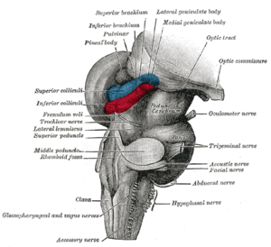
 後脳と中脳;後外側から見た図(外側膝状体は一番上にマークされている) | |
| 名称 | |
| 日本語 | 外側膝状体 |
| 英語 | lateral geniculate nucleus |
| ラテン語 | nucleus geniculatus lateralis |
| 略号 | LGN, LGB, LG |
| 関連構造 | |
| 上位構造 | 視床 |
| 動脈 | 前脈絡叢動脈、後脈絡叢動脈 |
| 静脈 | 視床線状体静脈 |
| 画像 | |
| アナトモグラフィー | 三次元CG |
| Digital Anatomist |
視床 視放線 冠状断(黒質) 水平断(膝状体) 視放線 |
| 関連情報 | |
| Brede Database | 階層関係、座標情報 |
| NeuroNames | 関連情報一覧 |
| NIF | 総合検索 |
| MeSH | Geniculate+Bodies |
| グレイの解剖学 | 書籍中の説明(英語) |
| テンプレートを表示 | |
外側膝状体(がいそくしつじょうたい, lateral geniculate nucleus,LGN,lateral geniculate body,LGB)は、脳の視床領域の一部であり、中枢神経系の網膜から情報を受け取り、視覚情報の処理を行う。
LGNは網膜から視神経、視交叉、視索を通って直接情報を受け取る。一次視覚野に視放線を通して直接投射する。また、LGNには一次視覚野からのフィードバック入力も多く投射している。
網膜神経節細胞が軸索を伸ばし、視神経としてLGNに投射している。
構造
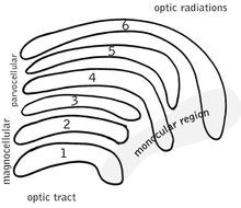
左右のLGNは固有の層構造を持つ。"geniculate"(膝状)とは"膝のように折れ曲がった"との意味。ヒトやマカクザルなど多くの霊長類では、LGNの各層のあいだに神経網(neruopil)があり、LGNニューロンの各層が"ケーキ"、神経網がアイシングとなって互いをはさみこむ、サンドイッチ状ないしはケーキ状の構造となっている。ヒトやマカクザルでは、LGNは大細胞層(magnocellular layer)が2層、小細胞層(parvocellular layer)が4層の、6層構造を持つとされる。こうした層の数は、霊長類の種によって異なり、種によっては別の層が加わることもある。
M, P, K 細胞
| 種類 | 大きさ | 機能 | 位置 |
| M: 大細胞(Magnocellular cells) | 大きな細胞体 | 情報処理に要する時間は短い。高速な処理ができるが、詳細な処理はできない | 1層と2層 |
| P: 小細胞(Parvocellular cells, "parvicellular"とも) | 小さい細胞体 | 情報処理に要する時間は長い。低速な処理であるが、詳細な処理が可能である。たとえば、P細胞は色の情報を処理できる。M細胞はこうした処理はおこなわない。 | 3, 4, 5層と6層 |
| K: 顆粒細胞(Koniocellular cells, "interlaminar"とも) | 非常に小さな細胞体 | M細胞やP細胞ほどは広く知られていない。 層のあいだに位置する。K細胞は機能的・神経化学的にM細胞やP細胞とは異なり、視覚野への第三のチャンネルとなっている。K細胞系が視知覚において果たす役割は、現在のところよく分かっていない。しかし、視覚と体性感覚・固有感覚情報の連合や、色覚への関与などが提案されている。 | M層とP層の層間 |

大細胞、小細胞、顆粒細胞の層は、類似する名称の神経節細胞と対応している。
小細胞層と大細胞層の神経線維は、以前はUngerleider-Mishkinの腹側系と背側系に対応すると考えられていた。しかし、近年の研究では、2つの処理経路は両者の神経線維をともに含んでいることが示されている[1]。
他の重要な網膜-皮質経路として網膜視蓋路がある。これは、上丘や視床枕核を経て、後頭頂葉や内側側頭葉へ至る経路である。
同側と反対側
M細胞とP細胞の分類に加えて、層は以下のように分類される:[2]
- LGNと同側(ipsilateral)の目は第2, 3, 5層に情報を送る
- LGNと反対側(contralateral)の目は第1, 4, 6層に情報を送る。
英語での簡単な記憶法は、"See I? I see, I see"を覚えることである。ここで、"see"は"contralateral"のCを表し、"I"は"ipsilateral"のIを表す。
こうした記述は、多くの霊長類のLGNに当てはまるが、全てではない。同側と反対側の目からこのように情報を受け取る方法は、メガネザルでは異なっている[3]。神経科学者には、このように一見するとメガネザルと他の霊長類に差があるように見えることが、メガネザルが比較的昔に生じた種であり、霊長類の進化とは独立だったことを示す、と考えるものもある。[4]。
視覚において、右側の目は右視野だけでなく、左視野の情報も受け取ることは、注意するべきである。このことは、左目を閉じれば分かる:右目によって、視野の右側も左側も見えるだろう。ただし、左視野の一部は、鼻によって遮蔽されている。
LGNでは、左右の目で対応する視野位置の情報は"積み重なって"おり、クラブサンドイッチの第1層から6層までトゥースピックで貫いたとすると、同じ視野位置を6回貫通することになる。
LGNへの入力
LGNは網膜からの入力を受ける。
いくつかの生物種では、LGNは視蓋(哺乳類での上丘)からの入力も受ける[5]。
LGNの出力
LGNから出力される情報は視放線として、内包のレンズ核後部を形成する。
LGNを出た軸索は、V1へと向かう。大細胞層1-2層と小細胞層3-6層は、ともにV1の4層にシナプスを作り、4cβは小細胞層から、4cαは大細胞層から入力を受ける。しかし、顆粒細胞層(1-6層の層間)は、V1の第2,3層へ軸索を送る。V1の6層からはLGNへフィードバックする軸索が送られる。
盲視の研究により、LGNからの神経投射はV1だけではなく高次の視覚領野にも及ぶことが示唆されている。盲視の患者はある視野位置での知覚を行うことができないが、こうした患者にテストを行うと、視野内の情報が無意識的にコードされていることが示される。このことは、LGNからの神経線維が、一次視覚野と高次視覚野の双方に投射することを示すと考えられることがある。
視覚における機能
LGNの機能はよく分かっていない。網膜は中心周辺拮抗処理により空間的無相関化(spatial decorrelation)を行うのに対して、LGNは時間的無相関化を行うと考えられることがある。こうした時空間無相関化により、より効率的なコーディングが可能になる。しかし、それ以上の処理が行われていることは確実だと考えられている。
視床の他の領域、特に他の中継核(relay nuclei)と同じように、LGNは視覚系が注意の焦点を特定の重要な情報に向けるのに役立つとされる。このことは、体の少し左側で音が聞こえたとすると、聴覚系が視覚系に対して、視覚的注意を空間のその領域に向けるよう、LGNを通じて"伝える"というものである。
LGNでは受容野がより精細化される場所でもある。
近年のヒトfMRI研究によると、空間的注意とサッカード眼球運動の双方がLGNの活動を修飾するとされる。
他の画像
| ウィキメディア・コモンズには、外側膝状体に関連するカテゴリがあります。 |

視床

脳幹の解剖。側面から見た図

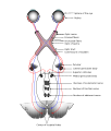
視神経と視索の走行を示す模式図

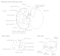
視床核

視索の3D模式図
脚注
| [脚注の使い方] |
- ^ Goodale & Milner, 1993, 1995.
- ^ Nicholls J., et. al. From Neuron to Brain: Fourth Edition. Sinauer Associates, Inc. 2001.
- ^ Rosa MG, Pettigrew JD, Cooper HM (1996) Unusual pattern of retinogeniculate projections in the controversial primate Tarsius. Brain Behav Evol 48(3):121-129.
- ^ Collins CE, Hendrickson A, Kaas JH (2005) Overview of the visual system of Tarsius. Anat Rec A Discov Mol Cell Evol Biol 287(1):1013-1025.
- ^ In Chapter 7, section "The Parcellation Hypothesis" of "Principals of Brain Evolution", Georg Striedter (Sinauer Associates, Sunderland, MA, USA, 2005)によると"...we now know that the LGN receives at least some inputs from the optic tectum (or superior colliculus) in many amniotes". 著者は"Wild, J.M. 1989. Pretectal and tectal projections to the homolog of the dorsal lateral geniculate nucleus in the pigeon - an anterograde and retrograde tracing study with cholera-toxin conjugated to horseradish-peroxidase. Brain Res 489: 130-137" および "Kaas, J.H., and Huerta, M.F. 1988. The subcortical visual system of primates. In: Steklis H. D., Erwin J., editors. Comparative primate biology, vol 4: neurosciences. New York: Alan Liss, pp. 327-391."を引用している。
外部リンク(英語)
- Blohm G and Schreiber C. LGN in the visual pathway. Retrieved September 1, 2004.
- Malpeli J. Malpeli Lab Home Page. Retrieved September 1, 2004.
| 典拠管理: 科学データベース |
|
|---|
wiki en
| Lateral geniculate nucleus | |
|---|---|
 Hind- and mid-brains; postero-lateral view. (Lateral geniculate body visible near top.) | |
| Details | |
| Part of | Thalamus |
| System | Visual |
| Artery | Anterior choroidal and Posterior cerebral |
| Vein | Terminal vein |
| Identifiers | |
| Latin | Corpus geniculatum laterale |
| Acronym(s) | LGN |
| NeuroNames | 352 |
| NeuroLex ID | birnlex_1662 |
| TA98 | A14.1.08.302 |
| TA2 | 5666 |
| FMA | 62209 |
| Anatomical terms of neuroanatomy [edit on Wikidata] | |
In neuroanatomy, the lateral geniculate nucleus (LGN; also called the lateral geniculate body or lateral geniculate complex) is a structure in the thalamus and a key component of the mammalian visual pathway. It is a small, ovoid, ventral projection of the thalamus where the thalamus connects with the optic nerve. There are two LGNs, one on the left and another on the right side of the thalamus. In humans, both LGNs have six layers of neurons (grey matter) alternating with optic fibers (white matter).
The LGN receives information directly from the ascending retinal ganglion cells via the optic tract and from the reticular activating system. Neurons of the LGN send their axons through the optic radiation, a direct pathway to the primary visual cortex. In addition, the LGN receives many strong feedback connections from the primary visual cortex.[1] In humans as well as other mammals, the two strongest pathways linking the eye to the brain are those projecting to the dorsal part of the LGN in the thalamus, and to the superior colliculus.[2]
Structure

Both the left and right hemisphere of the brain have a lateral geniculate nucleus, named after its resemblance to a bent knee (genu is Latin for "knee"). In humans as well as in many other primates, the LGN has layers of magnocellular cells and parvocellular cells that are interleaved with layers of koniocellular cells. In humans the LGN is normally described as having six distinctive layers. The inner two layers, (1 and 2) are magnocellular layers, while the outer four layers, (3,4,5 and 6), are parvocellular layers. An additional set of neurons, known as the koniocellular layers, are found ventral to each of the magnocellular and parvocellular layers.[3]: 227ff [4] This layering is variable between primate species, and extra leafleting is variable within species.
M, P, K cells
| Type | Size* | Source / Type of Information | Location | Response | Number |
| M: Magnocellular cells | Large | Rods; necessary for the perception of movement, depth, and small differences in brightness | Layers 1 and 2 | rapid and transient | ? |
| P: Parvocellular cells (or "parvicellular") | Small | Cones; long- and medium-wavelength ("red" and "green" cones); necessary for the perception of color and form (fine details). | Layers 3, 4, 5 and 6 | slow and sustained | ? |
| K: Koniocellular cells (or "interlaminar") | Very small cell bodies | Short-wavelength "blue" cones. | Between each of the M and P layers |

*Size relates to cell body, dendritic tree and receptive field
The magnocellular, parvocellular, and koniocellular layers of the LGN correspond with the similarly named types of retinal ganglion cells. Retinal P ganglion cells send axons to a parvocellular layer, M ganglion cells send axons to a magnocellular layer, and K ganglion cells send axons to a koniocellular layer.[5]: 269
Koniocellular cells are functionally and neurochemically distinct from M and P cells and provide a third channel to the visual cortex. They project their axons between the layers of the lateral geniculate nucleus where M and P cells project. Their role in visual perception is presently unclear; however, the koniocellular system has been linked with the integration of somatosensory system-proprioceptive information with visual perception, and it may also be involved in color perception.[6]
The parvo- and magnocellular fibers were previously thought to dominate the Ungerleider–Mishkin ventral stream and dorsal stream, respectively. However, new evidence has accumulated showing that the two streams appear to feed on a more even mixture of different types of nerve fibers.[7]
The other major retino–cortical visual pathway is the tectopulvinar pathway, routing primarily through the superior colliculus and thalamic pulvinar nucleus onto posterior parietal cortex and visual area MT.
Ipsilateral and contralateral layers
Layer 1, 2
- Large cells, called magnocellular pathways
- Input from Y-ganglion cells
- Very rapid conduction
- Colour blind system
Layer 3–6
- Parvocellular
- Input from X- ganglion cells
- Colour vision
- Moderate velocity.
Both the LGN in the right hemisphere and the LGN in the left hemisphere receive input from each eye. However, each LGN only receives information from one half of the visual field. This occurs due to axons of the ganglion cells from the inner halves of the retina (the nasal sides) decussating (crossing to the other side of the brain) through the optic chiasma (khiasma means "cross-shaped"). The axons of the ganglion cells from the outer half of the retina (the temporal sides) remain on the same side of the brain. Therefore, the right hemisphere receives visual information from the left visual field, and the left hemisphere receives visual information from the right visual field. Within one LGN, the visual information is divided among the various layers as follows:[8]
- the eye on the same side (the ipsilateral eye) sends information to layers 2, 3 and 5
- the eye on the opposite side (the contralateral eye) sends information to layers 1, 4 and 6.
This description applies to the LGN of many primates, but not all. The sequence of layers receiving information from the ipsilateral and contralateral (opposite side of the head) eyes is different in the tarsier.[9] Some neuroscientists suggested that "this apparent difference distinguishes tarsiers from all other primates, reinforcing the view that they arose in an early, independent line of primate evolution".[10]
In visual perception, the right eye gets information from the right side of the world (the right visual field), as well as the left side of the world (the left visual field). You can confirm this by covering your left eye: the right eye still sees to your left and right, although on the left side your field of view may be partially blocked by your nose.
Input
The LGN receives input from the retina and many other brain structures, especially visual cortex.
The principal neurons in the LGN receive strong inputs from the retina. However, the retina only accounts for a small percentage of LGN input. As much as 95% of input in the LGN comes from the visual cortex, superior colliculus, pretectum, thalamic reticular nuclei, and local LGN interneurons. Regions in the brainstem that are not involved in visual perception also project to the LGN, such as the mesencephalic reticular formation, dorsal raphe nucleus, periaqueuctal grey matter, and the locus coeruleus.[11] The LGN also receives some inputs from the optic tectum (also known as the superior colliculus).[12] These non-retinal inputs can be excitatory, inhibitory, or modulatory.[11]
Output
Information leaving the LGN travels out on the optic radiations, which form part of the retrolenticular portion of the internal capsule.
The axons that leave the LGN go to V1 visual cortex. Both the magnocellular layers 1–2 and the parvocellular layers 3–6 send their axons to layer 4 in V1. Within layer 4 of V1, layer 4cβ receives parvocellular input, and layer 4cα receives magnocellular input. However, the koniocellular layers, intercalated between LGN layers 1–6 send their axons primarily to the cytochrome-oxidase rich blobs of layers 2 and 3 in V1.[13] Axons from layer 6 of visual cortex send information back to the LGN.
Studies involving blindsight have suggested that projections from the LGN travel not only to the primary visual cortex but also to higher cortical areas V2 and V3. Patients with blindsight are phenomenally blind in certain areas of the visual field corresponding to a contralateral lesion in the primary visual cortex; however, these patients are able to perform certain motor tasks accurately in their blind field, such as grasping. This suggests that neurons travel from the LGN to both the primary visual cortex and higher cortex regions.[14]
Function in visual perception
This article needs additional citations for verification. Please help improve this article by adding citations to reliable sources. Unsourced material may be challenged and removed. Find sources: "Lateral geniculate nucleus" – news · newspapers · books · scholar · JSTOR (August 2019) (Learn how and when to remove this template message) |
The output of the LGN serves several functions.
Computations are achieved to determine the position of every major element in object space relative to the principal plane. Through subsequent motion of the eyes, a larger stereoscopic mapping of the visual field is achieved.[15]
It has been shown that while the retina accomplishes spatial decorrelation through center surround inhibition, the LGN accomplishes temporal decorrelation.[16] This spatial–temporal decorrelation makes for much more efficient coding. However, there is almost certainly much more going on.
Like other areas of the thalamus, particularly other relay nuclei, the LGN likely helps the visual system focus its attention on the most important information. That is, if you hear a sound slightly to your left, the auditory system likely "tells" the visual system, through the LGN via its surrounding peri-reticular nucleus, to direct visual attention to that part of space.[17] The LGN is also a station that refines certain receptive fields.[18]
Axiomatically determined functional models of LGN cells have been determined by Lindeberg [19][20] in terms of Laplacian of Gaussian kernels over the spatial domain in combination with temporal derivatives of either non-causal or time-causal scale-space kernels over the temporal domain. It has been shown that this theory both leads to predictions about receptive fields with good qualitative agreement with the biological receptive field measurements performed by DeAngelis et al.[21][22] and guarantees good theoretical properties of the mathematical receptive field model, including covariance and invariance properties under natural image transformations.[23] Specifically according to this theory, non-lagged LGN cells correspond to first-order temporal derivatives, whereas lagged LGN cells correspond to second-order temporal derivatives.
For an extensive overview of the function of the LGN in visual perception, see Ghodrati et al.[24]
Rodents
In rodents, the lateral geniculate nucleus contains the dorsal lateral geniculate nucleus (dLGN), the ventral lateral geniculate nucleus (vLGN), and the region in between called the intergeniculate leaflet (IGL). These are distinct subcortical nuclei with differences in function.
dLGN
The dorsolateral geniculate nucleus is the main division of the lateral geniculate body. The majority of input to the dLGN comes from the retina. It is laminated and shows retinotopic organization.[25]
vLGN
The ventrolateral geniculate nucleus has been found to be relatively large in several species such as lizards, rodents, cows, cats, and primates.[26] An initial cytoarchitectural scheme, which has been confirmed in several studies, suggests that the vLGN is divided into two parts. The external and internal divisions are separated by a group of fine fibers and a zone of thinly dispersed neurons. Additionally, several studies have suggested further subdivisions of the vLGN in other species.[27] For example, studies indicate that the cytoarchitecture of the vLGN in the cat differs from rodents. Although five subdivisions of the vLGN in the cat have been identified by some,[28] the scheme that divides the vLGN into three regions (medial, intermediate, and lateral) has been more widely accepted.
IGL
The intergeniculate leaflet is a relatively small area found dorsal to the vLGN. Earlier studies had referred to the IGL as the internal dorsal division of the vLGN. Several studies have described homologous regions in several species, including humans.[29]
The vLGN and IGL appear to be closely related based on similarities in neurochemicals, inputs and outputs, and physiological properties.
The vLGN and IGL have been reported to share many neurochemicals that are found concentrated in the cells, including neuropeptide Y, GABA, encephalin, and nitric oxide synthase. The neurochemicals serotonin, acetylcholine, histamine, dopamine, and noradrenaline have been found in the fibers of these nuclei.
Both the vLGN and IGL receive input from the retina, locus coreuleus, and raphe. Other connections that have been found to be reciprocal include the superior colliculus, pretectum, and hypothalamus, as well as other thalamic nuclei.
Physiological and behavioral studies have shown spectral-sensitive and motion-sensitive responses that vary with species. The vLGN and IGL seem to play an important role in mediating phases of the circadian rhythms that are not involved with light, as well as phase shifts that are light-dependent.[27]
Additional images

Thalamus

Dissection of brain-stem. Lateral view.

Scheme showing central connections of the optic nerves and optic tracts.

Thalamic nuclei

3D schematic representation of optic tracts
Brainstem. Posterior view.
References
- ^ Cudeiro, Javier; Sillito, Adam M. (2006). "Looking back: corticothalamic feedback and early visual processing". Trends in Neurosciences. 29 (6): 298–306. CiteSeerX 10.1.1.328.4248. doi:10.1016/j.tins.2006.05.002. PMID 16712965. S2CID 6301290.
- ^ Goodale, M. & Milner, D. (2004)Sight unseen.Oxford University Press, Inc.: New York.
- ^ Brodal, Per (2010). The central nervous system : structure and function (4th ed.). New York: Oxford University Press. ISBN 978-0-19-538115-3.
- ^ Carlson, Neil R. (2007). Physiology of behavior (9th ed.). Boston: Pearson/Allyn & Bacon. ISBN 978-0205467242.
- ^ Purves, Dale; Augustine, George; Fitzpatrick, David; Hall, William; Lamantia, Anthony-Samuel; White, Leonard (2011). Neuroscience (5. ed.). Sunderland, Mass.: Sinauer. ISBN 978-0878936953.
- ^ White, BJ; Boehnke, SE; Marino, RA; Itti, L; Munoz, DP (Sep 30, 2009). "Color-related signals in the primate superior colliculus". The Journal of Neuroscience. 29 (39): 12159–66. doi:10.1523/JNEUROSCI.1986-09.2009. PMC 6666157. PMID 19793973.
- ^ Goodale & Milner, 1993, 1995.
- ^ Nicholls J., et al. From Neuron to Brain: Fourth Edition. Sinauer Associates, Inc. 2001.
- ^ Rosa, MG; Pettigrew, JD; Cooper, HM (1996). "Unusual pattern of retinogeniculate projections in the controversial primate Tarsius". Brain, Behavior and Evolution. 48 (3): 121–9. doi:10.1159/000113191. PMID 8872317.
- ^ Collins, CE; Hendrickson, A; Kaas, JH (Nov 2005). "Overview of the visual system of Tarsius". The Anatomical Record Part A: Discoveries in Molecular, Cellular, and Evolutionary Biology. 287 (1): 1013–25. doi:10.1002/ar.a.20263. PMID 16200648.
- ^ a b Guillery, R; SM Sherman (Jan 17, 2002). "Thalamic relay functions and their role in corticocortical communication: generalizations from the visual system". Neuron. 33 (2): 163–75. doi:10.1016/s0896-6273(01)00582-7. PMID 11804565.
- ^ In Chapter 7, section "The Parcellation Hypothesis" of "Principles of Brain Evolution", Georg F. Striedter (Sinauer Associates, Sunderland, MA, USA, 2005) states, "...we now know that the LGN receives at least some inputs from the optic tectum (or superior colliculus) in many amniotes". He cites "Wild, J.M. (1989). "Pretectal and tectal projections to the homolog of the dorsal lateral geniculate nucleus in the pigeon—an anterograde and retrograde tracing study with cholera-toxin conjugated to horseradish-peroxidase". Brain Res. 479 (1): 130–137. doi:10.1016/0006-8993(89)91342-5. PMID 2924142. S2CID 29034684." and also "Kaas, J.H., and Huerta, M.F. 1988. The subcortical visual system of primates. In: Steklis H. D., Erwin J., editors. Comparative primate biology, vol 4: neurosciences. New York: Alan Liss, pp. 327–391.
- ^ Hendry, Stewart H. C.; Reid, R. Clay (2000). "The koniocellular pathway in primate vision". Annual Review of Neuroscience. 23: 127–153. doi:10.1146/annurev.neuro.23.1.127. PMID 10845061.
- ^ Schmid, Michael C.; Mrowka, Sylwia W.; Turchi, Janita; et al. (2010). "Blindsight depends on the lateral geniculate nucleus". Nature. 466 (7304): 373–377. Bibcode:2010Natur.466..373S. doi:10.1038/nature09179. PMC 2904843. PMID 20574422.
- ^ Lindstrom, S. & Wrobel, A. (1990) Intracellular recordings from binocularly activated cells in the cats dorsal lateral geniculate nucleus Acta Neurobiol Exp vol 50, pp 61–70
- ^ Dawei W. Dong and Joseph J. Atick, Network–Temporal Decorrelation: A Theory of Lagged and Nonlagged Responses in the Lateral Geniculate Nucleus, 1995, pp. 159–178.
- ^ McAlonan, K.; Cavanaugh, J.; Wurtz, R. H. (2006). "Attentional Modulation of Thalamic Reticular Neurons". Journal of Neuroscience. 26 (16): 4444–4450. doi:10.1523/JNEUROSCI.5602-05.2006. PMC 6674014. PMID 16624964.
- ^ Tailby, C.; Cheong, S. K.; Pietersen, A. N.; Solomon, S. G.; Martin, P. R. (2012). "Colour and pattern selectivity of receptive fields in superior colliculus of marmoset monkeys". The Journal of Physiology. 590 (16): 4061–4077. doi:10.1113/jphysiol.2012.230409. PMC 3476648. PMID 22687612.
- ^ Lindeberg, T. (2013). "A computational theory of visual receptive fields". Biological Cybernetics. 107 (6): 589–635. doi:10.1007/s00422-013-0569-z. PMC 3840297. PMID 24197240.
- ^ Lindeberg, T. (2021). "Normative theory of visual receptive fields". Heliyon. 7 (1): e05897. doi:10.1016/j.heliyon.2021.e05897. PMC 7820928. PMID 33521348.
- ^ DeAngelis, G. C.; Ohzawa, I.; Freeman, R. D. (1995). "Receptive field dynamics in the central visual pathways". Trends Neurosci. 18 (10): 451–457. doi:10.1016/0166-2236(95)94496-r. PMID 8545912. S2CID 12827601.
- ^ G. C. DeAngelis and A. Anzai "A modern view of the classical receptive field: linear and non-linear spatio-temporal processing by V1 neurons. In: Chalupa, L.M., Werner, J.S. (eds.) The Visual Neurosciences, vol. 1, pp. 704–719. MIT Press, Cambridge, 2004.
- ^ Lindeberg, T. (2013). "Invariance of visual operations at the level of receptive fields". PLOS ONE. 8 (7): e66990. arXiv:1210.0754. Bibcode:2013PLoSO...866990L. doi:10.1371/journal.pone.0066990. PMC 3716821. PMID 23894283.
- ^ M. Ghodrati, S.-M. Khaligh-Razavi, S.R. Lehky, Towards building a more complex view of the lateral geniculate nucleus: recent advances in understanding its role, Prog. Neurobiol. 156:214–255, 2017.
- ^ Grubb, Matthew S.; Francesco M. Rossi; Jean-Pierre Changeux; Ian D. Thompson (Dec 18, 2003). "Abnormal functional organization in the dorsal lateral geniculate nucleus of mice lacking the beta2 subunit of the nicotinic acetylcholine receptor". Neuron. 40 (6): 1161–1172. doi:10.1016/s0896-6273(03)00789-x. PMID 14687550.
- ^ Cooper, H.M.; M. Herbin; E. Nevo (Oct 9, 2004). "Visual system of a naturally microphthalamic mammal: The blind mole rat, Spalax ehrenbergl". Journal of Comparative Neurology. 328 (3): 313–350. doi:10.1002/cne.903280302. PMID 8440785. S2CID 28607983.
- ^ a b Harrington, Mary (1997). "The ventral lateral geniculate nucleus and the intergeniculate leaflet: interrelated structures in the visual and circadian systems". Neuroscience and Biobehavioral Reviews. 21 (5): 705–727. doi:10.1016/s0149-7634(96)00019-x. PMID 9353800. S2CID 20139828.
- ^ Jordan, J.; H. Hollander (1972). "The structure of the ventral part of the lateral geniculate nucleus – a cyto- and myeloarchitectonic study in the cat". Journal of Computational Neuroscience. 145 (3): 259–272. doi:10.1002/cne.901450302. PMID 5030906. S2CID 30586321.
- ^ Moore, Robert Y. (1989). "The geniculohypothalamic tract in monkey and man". Brain Research. 486 (1): 190–194. doi:10.1016/0006-8993(89)91294-8. PMID 2720429. S2CID 33543381.
External links
- Malpeli J. Malpeli Lab Home Page. Retrieved September 1, 2004.
- Stained brain slice images which include the "lateral%20geniculate%20nucleus" at the BrainMaps project
- Atlas image: eye_38 at the University of Michigan Health System – "The Visual Pathway from Below"
- Stained brain slice images which include the "lgn" at the BrainMaps project
- MedicalMnemonics.com: 307 640
Optical illusions (list) | ||
|---|---|---|
| Illusions |
| |
| Popular culture |
| |
| Related |
| |
Anatomy of the diencephalon of the human brain | |||||||||||||||
|---|---|---|---|---|---|---|---|---|---|---|---|---|---|---|---|
| Epithalamus |
| ||||||||||||||
| Thalamus |
| ||||||||||||||
| Hypothalamus |
| ||||||||||||||
| Subthalamus |
| ||||||||||||||
The cranial nerves | |||||||||||||
|---|---|---|---|---|---|---|---|---|---|---|---|---|---|
| Terminal (CN 0) |
| ||||||||||||
| Olfactory (CN I) |
| ||||||||||||
| Optic (CN II) |
| ||||||||||||
| Oculomotor (CN III) |
| ||||||||||||
| Trochlear (CN IV) |
| ||||||||||||
| Trigeminal (CN V) |
| ||||||||||||
| Abducens (CN VI) |
| ||||||||||||
| Facial (CN VII) |
| ||||||||||||
| Vestibulocochlear (CN VIII) |
| ||||||||||||
| Glossopharyngeal (CN IX) |
| ||||||||||||
| Vagus (CN X) |
| ||||||||||||
| Accessory (CN XI) |
| ||||||||||||
| Hypoglossal (CN XII) |
| ||||||||||||
| Authority control: Scientific databases |
|
|---|
English Journal
- Reciprocal inhibition and slow calcium decay in perigeniculate interneurons explain changes of spontaneous firing of thalamic cells caused by cortical inactivation.
- Rogala J, Waleszczyk WJ, Lęski S, Wróbel A, Wójcik DK.SourceDepartment of Neurophysiology, Nencki Institute of Experimental Biology, 3 Pasteur St, 02-093, Warsaw, Poland.
- Journal of computational neuroscience.J Comput Neurosci.2013 Jun;34(3):461-76. doi: 10.1007/s10827-012-0430-8. Epub 2012 Nov 13.
- The role of cortical feedback in the thalamocortical processing loop has been extensively investigated over the last decades. With an exception of several cases, these searches focused on the cortical feedback exerted onto thalamo-cortical relay (TC) cells of the dorsal lateral geniculate nucleus (L
- PMID 23150147
- Responses of primate LGN cells to moving stimuli involve a constant background modulation by feedback from area MT.
- Jones HE, Andolina IM, Grieve KL, Wang W, Salt TE, Cudeiro J, Sillito AM.SourceDepartment of Visual Neuroscience, UCL Inst. of Ophthalmology, London, United Kingdom. Electronic address: hjones@ioores.co.uk.
- Neuroscience.Neuroscience.2013 May 2. pii: S0306-4522(13)00388-6. doi: 10.1016/j.neuroscience.2013.04.055. [Epub ahead of print]
- The feedback connections from the cortical motion area MT, to layer 6 of the primary visual cortex (V1), have the capacity to drive a cascaded feedback influence from the layer 6 cortico-geniculate cells back to the lateral geniculate nucleus (LGN) relay cells. This introduces the possibility of a r
- PMID 23644057
- Crystal structure of the guanylate kinase domain from discs large homolog 1 (DLG1/SAP97).
- Mori S, Tezuka Y, Arakawa A, Handa N, Shirouzu M, Akiyama T, Yokoyama S.SourceRIKEN Systems and Structural Biology Center, Tsurumi-ku, Yokohama 230-0045, Japan; Department of Biophysics and Biochemistry, Graduate School of Science, The University of Tokyo, Bunkyo-ku, Tokyo 113-0033, Japan.
- Biochemical and biophysical research communications.Biochem Biophys Res Commun.2013 Apr 25. pii: S0006-291X(13)00695-5. doi: 10.1016/j.bbrc.2013.04.056. [Epub ahead of print]
- Discs large homolog 1 (DLG1/SAP97) is involved in the development and regulation of neuronal and immunological synapses. DLG1 is a member of the membrane associated guanylate kinase (MAGUK) family of proteins, which function as molecular scaffolds. The C-terminal guanylate kinase (GK) domain of DLG1
- PMID 23624197
Japanese Journal
- 〈Original Papers〉網膜-外側膝状体間における視覚情報のリマッピング
- 近畿大学生物理工学部紀要 = Memoirs of the Faculty of Biology-Oriented Science and Technology of Kindai University (38), 11-20, 2016-10-31
- NAID 120005983716
- Interphase adhesion geometry is transmitted to an internal regulator for spindle orientation via caveolin-1
- Nature Communications 7, 2016-06-13
- NAID 120005774665
- 自由行動下ラット視床外側膝状体単一神経細胞の慢性記録実験
- 京都産業大学総合学術研究所所報 10, 39-47, 2015-07
- NAID 110009921176
Related Links
- 最低のお金で最高の旅を!ラグナトラベル 発券するまで変更、キャンセルは無料!業務渡航にも最適。 スタッフ全員オーストラリア在住経験ありなのでオーストラア旅行 ワーキングホリデー 留学のご相談もどうぞ!
- The latest from LGNサクセスサポート (@LGN_777). 309$の1回きりの投資で世界230カ国に展開可能なインターネットビジネスであるLGNを強力に親切にサポートしております。 ぜひ、LGN紹介ホームページを覗いてください。 日本上陸前 ...
Related Pictures


★リンクテーブル★
| リンク元 | 「ループス腎炎」「外側膝状核」「外側膝状体核」 |
| 関連記事 | 「L」「LG」 |
「ループス腎炎」
概念
- 全身性工リテマトーデスは、自己免疫反応の結果生じる免疫復合体が全身の血管に沈着して種々の病像を呈する疾患
- この免疫複合体が腎臓に沈着、あるいは局所で産生されることにより惹起される腎炎。免疫複合体型腎炎
疫学
- SLEの60-80%にループス腎炎が合併し、その約半数がネフローゼ症候群に移行する。
- SLE全体の予後を決定するのは、中枢神経系の病変、肺病変(肺出血など)、重症の溶血貧血/血小板減少症、全身性血管炎、ないしループス腎炎。
病理
組織分類
I型 :正常糸球体 normal
II型 :メサンギウム増殖性糸球体腎炎 mesangial alterations
III型:巣状分節状糸球体腎炎 focal GN
IV型 :びまん性増殖性糸球体腎炎 diffuse proliferative GN
V型 :びまん性膜性糸球体腎炎 diffuse membranous GN
VI型 :硬化性糸球体腎炎 advanced sclerosing GN
治療
- 1. 副腎皮質ステロイド(特にパルス療法)
- 2. 副腎皮質ステロイドと免疫抑制薬の併用療法
- 3. 血漿交換療法
- 4. 抗凝固療法、血液透析
国試
「外側膝状核」
「外側膝状体核」
- 英
- ()
「L」
「LG」


