WordNet
- of or relating to or in the region of the mastoid process
- process of the temporal bone behind the ear at the base of the skull (同)mastoid_process, mastoid bone, mastoidal
- relating to or resembling a nipple (同)mastoidal
- any membranous gap between the bones of the cranium in an infant or fetus (同)fontanel, soft_spot
PrepTutorEJDIC
- 乳様突起の / 〈C〉乳様突起(耳の後ろの側頭骨にある)
Wikipedia preview
出典(authority):フリー百科事典『ウィキペディア(Wikipedia)』「2022/02/27 00:07:01」(JST)
wiki en
UpToDate Contents
全文を閲覧するには購読必要です。 To read the full text you will need to subscribe.
- 1. 小児における身体的診察:HEENT the pediatric physical examination heent
- 2. グラム陰性桿菌性髄膜炎:疫学、臨床的特徴、および診断 gram negative bacillary meningitis epidemiology clinical features and diagnosis
- 3. 小児における肺炎球菌性髄膜炎 pneumococcal meningitis in children
- 4. 急性運動失調を呈する小児に対するアプローチ approach to the child with acute ataxia
- 5. 小児における急性乳様突起炎:臨床的特徴および診断 acute mastoiditis in children clinical features and diagnosis
English Journal
- Accuracy of ultrasound in assessing cerebellar haemorrhages in very low birthweight babies.
- Parodi A1, Rossi A2, Severino M2, Morana G2, Sannia A1, Calevo MG3, Malova M1, Ramenghi LA1.
- Archives of disease in childhood. Fetal and neonatal edition.Arch Dis Child Fetal Neonatal Ed.2015 Jul;100(4):F289-92. doi: 10.1136/archdischild-2014-307176. Epub 2015 Jan 30.
- OBJECTIVE: To assess diagnostic accuracy of cranial ultrasound (CUS) performed through the anterior fontanelle (AF) and mastoid fontanelle (MF) in detecting cerebellar haemorrhages (CBH) in very low birthweight (VLBW) infants.SETTING: Third-level neonatal intensive care unit (NICU).DESIGN: VLBW infa
- PMID 25637005
- Study on the influence of the fetus head molding on the biomechanical behavior of the pelvic floor muscles, during vaginal delivery.
- Silva ME1, Oliveira DA1, Roza TH1, Brandão S2, Parente MP3, Mascarenhas T4, Natal Jorge RM1.
- Journal of biomechanics.J Biomech.2015 Jun 25;48(9):1600-5. doi: 10.1016/j.jbiomech.2015.02.032. Epub 2015 Feb 26.
- Pelvic floor injuries during vaginal delivery are considered a significant risk factor to develop pelvic floor dysfunction. The molding of the fetus head during vaginal delivery facilitates the labor progress, since it adjusts to the birth canal geometry. In this work, a finite element model was use
- PMID 25757665
- The Incidence and Topographic Distribution of Sutures Including Wormian Bones in Human Skulls.
- Cirpan S1, Aksu F, Mas N.
- The Journal of craniofacial surgery.J Craniofac Surg.2015 Jun 24. [Epub ahead of print]
- OBJECTIVE: The Wormian Bones are accessory bones located within the cranial sutures and fontanelles. The present article examines the incidence of Wormian Bones and compares the number and topographic distribution between the sutures including Wormian Bones in skulls of West Anatolian Population.MET
- PMID 26114515
Related Links
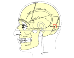
- mas·toid fon·ta·nelle [TA] the membranous interval on either side between the mastoid angle of the parietal bone, the petrous portion of the temporal bone, and the occipital bone. Synonym(s): fonticulus mastoideus [TA], fonticulus ...
- The mastoid fontanelle is one of two areas of an infant's head where the skull bones have not completely covered the brain. It is also known as one of the soft spots. There is one mastoid fontanelle on each side of the head, just ...
★リンクテーブル★
| リンク元 | 「泉門」「後側頭泉門」 |
| 関連記事 | 「mastoid」 |
「泉門」
- 英
- fontanelle, fontanelles
- ラ
- fonticulus, fonticuli cranii
- 同
- 頭蓋泉門
- 関
- [[]]
泉門
「後側頭泉門」
- 英
- mastoid fontanelle (N,KA), mastoid fontanel (HT)
- 関
- 泉門
- 側頭骨-後頭骨間、さらに上方では後頭骨によって囲まれた泉門
「mastoid」
