WordNet
- a missing piece (as a gap in a manuscript)
PrepTutorEJDIC
- 《文》すきま,割れ目 / 欠文,脱字 / 《文》(時間などの)中絶,とぎれ
Wikipedia preview
出典(authority):フリー百科事典『ウィキペディア(Wikipedia)』「2022/11/12 03:32:15」(JST)
wiki en
UpToDate Contents
全文を閲覧するには購読必要です。 To read the full text you will need to subscribe.
- 1. 鼻症状の原因:概要etiologies of nasal symptoms an overview [show details]
- 2. 裂孔ヘルニアhiatus hernia [show details]
- 3. 逆流性食道炎の病態生理pathophysiology of reflux esophagitis [show details]
- 4. component separation法(腹壁瘢痕ヘルニア修復法)の概要overview of component separation [show details]
- 5. 傍食道型食道裂孔ヘルニアへの外科的マネージメントsurgical management of paraesophageal hernia [show details]
English Journal
- The Anterior Ethmoidal "Genu": A Newly Appreciated Anatomic Landmark for Endoscopic Sinus Surgery.
- Bolger WE, Stammberger H, Ishii M, Ponikau J, Solaiyappan M, Zinreich SJ.
- Clinical anatomy (New York, N.Y.). 2019 May;32(4)534-540.
- Human sinonasal anatomy varies widely between patients, challenging surgeons operating in the sinuses. Ethmoid sinus anatomy is so variable it has been referred to as a labyrinth. Accordingly, reliable, consistent anatomic landmarks aid surgeons operating in this region. The goal of this investigati
- PMID 30719771
- Localization of the Maxillary Ostium in Relation to the Reduction of Depressed Nasomaxillary Fractures.
- Hwang K, Wu X, Kim H, Kang YH.
- The Journal of craniofacial surgery. 2018 Jul;29(5)1358-1362.
- The aim of this study was to elucidate the precise location of the maxillary ostium using computed tomography for the reduction of depressed nasomaxillary fractures.Computed tomography images (61 males, 42 females; age range, 3-97 years) were analyzed. Coronal sections were cut every 3 mm.The prim
- PMID 29521750
- Anatomic description of the middle meatus and classification of the hiatus semilunaris into five types based upon morphological characteristics.
- Dahlstrom K, Olinger A.
- Clinical anatomy (New York, N.Y.). 2014 Mar;27(2)176-81.
- Anatomical variation of the lateral nasal wall, including the pathway from the frontal, ethmoidal, and maxillary sinuses may affect the communication between the paranasal sinuses and the nasal cavity. The middle meatus and hiatus semilunaris are areas where variations can occur which predispose pat
- PMID 23836582
Japanese Journal
- CT前額断像によるOsteomeatal unit(OMU:洞口鼻道系)と歯性上顎洞炎の関連
- 歯科放射線 47(2), 47-52, 2007
- NAID 130004503190
- 小児慢性副鼻こう炎に対するせん刺洗浄療法 ステロイド加抗生剤注入の効果について:ステロイド加抗生剤注入の効果について
- 耳鼻咽喉科展望 28(1), 11-17, 1985
- NAID 130003794264
- ヒト胎児の蝸牛窓の発生について
- 耳鼻と臨床 28(5Supplement3), 845-849, 1982
- NAID 130004402716
Related Links
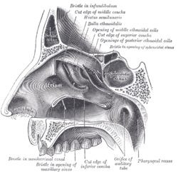
- The hiatus semilunaris is a semicircular shaped opening located on the lateral wall of the nasal cavity. It is a component of the ostiomeatal complex and serves as the opening for the frontal and maxillary sinuses and the anterior ethmoid air cells. It is inferior to the ethmoid bulla and the uncinate process forms its anterior border.
- The hiatus semilunaris (or semilunar hiatus) is a crescent-shaped groove in the lateral wall of the nasal cavity just inferior to the ethmoidal bulla. It is the location of the openings for the frontal sinus, maxillary sinus, and anterior ethmoidal sinus. It is bounded inferiorly and anteriorly by the sharp concave margin of the uncinate process of ...
- noun hiatus semi· lu· nar· is -ˌsem-i-lü-ˈnar-əs : a curved fissure in the nasal passages into which the frontal and maxillary sinuses open Dictionary Entries Near hiatus semilunaris hiatus hiatus semilunaris Hib See More Nearby Entries Cite this Entry Style “Hiatus semilunaris.”
★リンクテーブル★
| リンク元 | 「半月裂孔」 |
| 関連記事 | 「hiatus」 |
「半月裂孔」
- 英
- semilunar hiatus (Z)
- ラ
- hiatus semilunaris
- 同
- 半月状裂孔
- 図:N.45
「hiatus」
