Extrinsic muscles of the tongue. Left side. (Hyoglossus visible at center.)
Muscles of the neck. Anterior view. Hyoglossal muscle in purple
Details
Origin
Hyoid
Insertion
side of the tongue
Nerve
Hypoglossal (CN XII)
Actions
depresses and retracts the tongue
Identifiers
Latin
musculus hyoglossus
TA98
A05.1.04.102
TA2
2118
FMA
46691
Anatomical terms of muscle
[edit on Wikidata]
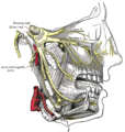
The hyoglossus, thin and quadrilateral, arises from the side of the body and from the whole length of the greater cornu of the hyoid bone, and passes almost vertically upward to enter the side of the tongue, between the styloglossus and the inferior longitudinal muscle of the tongue. It forms a part of the floor of submandibular triangle.
Contents
1Structure
2Function
3Additional images
4References
5External links
Structure
The fibers arising from the body of the hyoid bone overlap those from the greater cornu.
Structures that are medial/deep to the hyoglossus are the glossopharyngeal nerve (cranial nerve 9), the stylohyoid ligament and the lingual artery and lingual vein.
The lingual vein passes medial to the hyoglossus, and the lingual artery passes deep to the hyoglossus. Laterally, in between the hyoglossus muscle and the mylohyoid muscle lay several important structures (from upper to lower): sublingual gland, submandibular duct, lingual nerve, vena comitans of hypoglossal nerve, and the hypoglossal nerve. Note, posteriorly, the lingual nerve is superior to the submandibular duct and a portion of the submandibular salivary gland protrudes into the space between the hyoglossus and mylohyoid muscles.
Function
The hyoglossus depresses and retracts the tongue and makes the dorsum more convex.
Additional images
Hyoid bone. Anterior surface. Enlarged.
Muscles of the neck. Lateral view.
The internal carotid and vertebral arteries. Right side.
Distribution of the maxillary and mandibular nerves, and the submaxillary ganglion.
Hypoglossal nerve, cervical plexus, and their branches.
Coronal section of tongue, showing intrinsic muscles.
Hyoglossus Muscle
References
This article incorporates text in the public domain from page 1129 of the 20th edition of Gray's Anatomy (1918)
External links
Anatomy figure: 34:02-09 at Human Anatomy Online, SUNY Downstate Medical Center
"Anatomy diagram: 25420.000-1". Roche Lexicon - illustrated navigator. Elsevier. Archived from the original on 2013-12-31.
Diagram
v
t
e
Muscles of the head
Extraocular
Oblique
inferior
superior
Rectus
superior
inferior
medial
lateral
Levator palpebrae superioris
superior tarsal
Masticatory
Masseter
Temporalis
sphenomandibularis
Pterygoid
lateral
medial
Fascia
masseteric
temporal
Facial
Ear
Auricular
anterior
superior
posterior
Temporoparietalis
Scalp/eyelid
Occipitofrontalis
occipitalis
frontalis
Orbicularis oculi
depressor supercilii
Corrugator supercilii
Levator palpebrae superioris
Nose
Procerus
Nasalis
dilator naris
Depressor septi nasi
Levator labii superioris alaeque nasi
Mouth
Levator anguli oris
Levator labii superioris
Zygomaticus
major
minor
Orbicularis oris
Risorius
Buccinator
Depressor anguli oris
Depressor labii inferioris
Mentalis
Transversus menti
Soft palate
Veli palatini
tensor
levator
Musculus uvulae
Palatopharyngeus
Palatoglossus
Tongue
Extrinsic
Genioglossus
Hyoglossus
chondroglossus
Styloglossus
Palatoglossus
Intrinsic
Superior longitudinal
Inferior longitudinal
Transverse
Vertical
Portal:
Anatomy
Authority control: Scientific databases
Terminologia Anatomica
UpToDate Contents
全文を閲覧するには購読必要です。 To read the full text you will need to subscribe.
… The GGh pulls the tongue forward, while the GGo pulls the tongue down. Styloglossus (SG) and hyoglossus (HG) muscles, which are innervated by the lateral division of CN XII. The HG retracts and depresses …
English Journal
Anatomy of the lingual nerve: Application to oral surgery.
Shimotakahara R, Lee H, Mine K, Ogata S, Tamatsu Y.
Clinical anatomy (New York, N.Y.). 2019 Jul;32(5)635-641.
The purpose of this research is to obtain morphological information about the traveling route, branching pattern, and distribution within the tongue of the lingual nerve, all of which are important for oral surgical procedures. Using 20 sides from 10 Japanese cadaveric heads, we followed the lingual
Morphological Features of the Branching Pattern of the Hypoglossal Nerve.
, S S, Y Y, Y Y, h h, .
Anatomical record (Hoboken, N.J. : 2007). 2019 Apr;302(4)558-567.
The hypoglossal or twelfth cranial nerve is the motor nerve to the extrinsic and intrinsic muscles of the tongue, and the superior root of the ansa cervicalis and the thyrohyoid and geniohyoid branches are delivered through the nerve. This study investigated the muscular branches of the hypoglossal
Radiographic muscle invasion not a recurrence predictor in HPV-associated oropharyngeal squamous cell carcinoma.
Farzal Z, Du E, Yim E, Mazul A, Zevallos JP, Huang BY, Hackman TG.
The Laryngoscope. 2019 04;129(4)871-876.
To determine whether muscle invasion evident on pretreatment imaging in p16 + oropharyngeal squamous cell carcinoma (OPSCC) correlates with recurrence. Retrospective review. Two-hundred and seventy-six patients with p16 + OPSCC treated at a tertiary referral center from 2003 to 2015 were ana
Hyoglossus and genioglossus activity are synchronous with respiration, and activity of these muscles correlates well with increases in pharyngeal airway size during breathing (Brouillette & Bradley 1980, Mathew et al 1984a, ,
Note, posteriorly, the lingual nerve is superior to the submandibular duct and a portion of the submandibular salivary gland protrudes into the space between the hyoglossus and mylohyoid muscles. The hyoglossus depresses and retracts the tongue and makes the dorsum more convex. ・ quadrilateral : 四辺形の ・ greater cornu of the hyoid ...